SERVICES

If you are experiencing pain, we want to help! We are dedicated to helping patients regain pain-free lives. Oftentimes, people do not seek help for pain until it worsens. We want to help at the onset of symptoms, so they do not progress.
Before the injection procedure begins, topical anesthesia is applied to the skin. Next, in order to prevent healthy nerve roots from being exposed to too much medication, the physician will use imaging technology such as fluoroscopy to guide the insertion of the needle and to confirm its correct placement in the epidural space. In addition, contrast dye is typically injected in order to observe where the medication will be administered and to ensure that it will be properly distributed throughout the targets areas. The administration of steroids and an anesthetic such as Lidocaine directly onto the nerves roots results in dramatic or complete pain relief. The steroid decreases inflammation, while the anesthetic disrupts pain signal transmission.
A cervical epidural steroid injection is a non-surgical, outpatient procedure that can be quickly performed. Patients who receive epidural steroid injections typically experience an immediate decrease in their level of pain or complete pain relief.
A herniated disc: This occurs when one of the discs that act as a cushion between spinal vertebrae, ruptures, causing some of the jelly-like substance to leak out, irritating nearby nerves.
Cervical spondylosis: This relates to damage to spinal discs in the neck, as a result of age-related wear and tear.
Cervical radiculopathy: This refers to pain that results from any kind of condition or injury that causes damage to the nerves in the neck.
Cervical spinal stenosis: This is a narrowing of the open spaces in the spine that results in pressure on the spinal cord and nerves, causing symptoms such as pain and numbness.
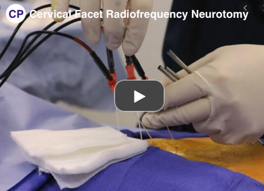
Radiofrequency neurotomy is usually done by a doctor who specializes in treating pain. The goal is to reduce chronic back, neck, hip or knee pain that hasn’t improved with medications or physical therapy, or when surgery isn’t an option. For example, your doctor may suggest the procedure if you have back pain that:
- Occurs on one or both sides of your lower back
- Spreads to the buttocks and thighs (but not below the knee)
- Feels worse if you twist or lift something
- Feels better when you’re lying down
Radiofrequency neurotomy is an outpatient procedure, so you’ll go home later that same day. The doctor will the use a special X-ray machine (fluoroscope) to guide the radiofrequency needles to the precise area — so only the targeted nerve tissue will be treated. During a radiofrequency neurotomy procedure, the heat comes from a small electrical current that travels through a needle that has been inserted next to these small nerves. The heat is directed at the nerve that is causing the pain, so that nearby healthy nerves are not damaged during the procedure.
The radiofrequency neurotomy procedure can help patients who suffer from:
- Osteoarthritis, a chronic condition of the joints in which the cartilage, the smooth material between the joints, wears away;
- Spine conditions that are a result of a traumatic injury, such as a car accident in which the spine is injured.
Your doctor may order an EMG if you have signs or symptoms that may indicate a nerve or muscle disorder. Such symptoms may include:
- Tingling
- Numbness
- Muscle weakness
- Muscle pain or cramping
- Certain types of limb pain
The results of an EMG can help your doctor determine the underlying cause of these symptoms. Possible causes could include:
- muscle disorders, such as muscular dystrophy
- disorders that affect the ability of the motor neuron to send electrical signals to the muscle, such as myasthenia gravis
- radiculopathies
- peripheral nerve disorders that affect the nerves outside the spinal cord, such as carpal tunnel syndrome
- nerve disorders, such as amyotrophic lateral sclerosis (ALS)
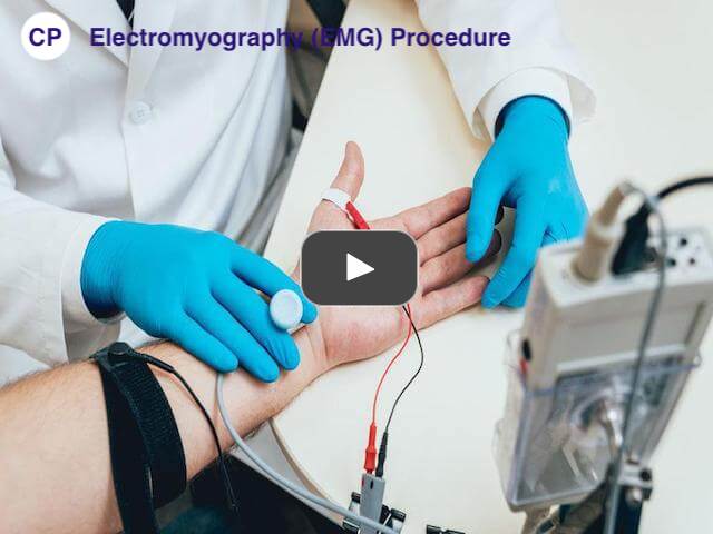
There are two components to an EMG test: the nerve conduction study and needle EMG. The nerve conduction study is the first part of the procedure. It involves placing small sensors called surface electrodes on the skin to assess the ability of the motor neurons to send electrical signals. The second part of the EMG procedure, known as needle EMG, also uses sensors to evaluate electrical signals. The sensors are called needle electrodes, and they’re directly inserted into muscle tissue to evaluate muscle activity when at rest and when contracted.
The nerve conduction study is performed first. During this portion of the procedure, your doctor will apply several electrodes to the surface of your skin, usually in the area where you’re experiencing symptoms. These electrodes will evaluate how well your motor neurons communicate with your muscles. Once the test is complete, the electrodes are removed from the skin. After the nerve conduction study, your doctor will perform the needle EMG. Your doctor will first clean the affected area with an antiseptic. Then, they will use a needle to insert electrodes into your muscle tissue. You may feel slight discomfort or pain while the needle is being inserted.
The needle electrodes will evaluate the electrical activity of your muscles when contracted and when at rest. These electrodes will be removed after the test is over. During both parts of the EMG procedure, the electrodes will deliver tiny electrical signals to your nerves. A computer will translate these signals into graphs or numerical values that can be interpreted by your doctor. The entire procedure should take between 30 and 60 minutes.
A facet joint injection is performed to treat neck and back pain in combination with other non-surgical spine treatments like rest, medications, chiropractic manipulations, and physical therapy. It is one of two types of injections that treat pain arising from the joints of the spine:
- Facet joint injections – place medication directly into the joint.
- Medial branch blocks (MBBs) – stop the transmission of pain signals by targeting the nerves along the facet joint.
This treatment does not offer permanent pain relief. Patients may have one or two repeat injections over a six-month period. It is not recommended that a patient receive more than three injections in this time frame.
In order to guide the injection to the facet joint, your pain specialist will use fluoroscopic (X-ray) guidance during the injection. This will allow the physician to see the joint and ensure that the needle is in place before injecting the anesthetic-cortisone mixture. The procedure takes between 5-15 minutes. Thanks to the anesthetic used, the injection can provide immediate pain relief.
We Provide the highest level of satisfaction
care & services to our patients.
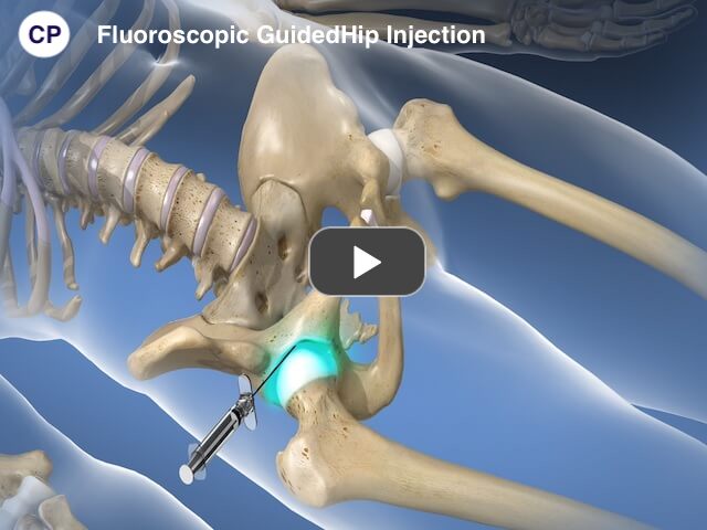
When you are in the procedure room, you will be asked to lie on your back on a cushioned x-ray table. A fluoroscope (x-ray machine) assists the provider in visualizing the hip. After visualization of the joint under x-ray, a small needle is placed into the skin, and positioned into the joint space. A small amount of a solution of local anesthetic (numbing medication) and a small amount of x-ray contrast is used to confirm placement into the joint. Once confirmed a cortisone derivative (anti-inflammatory medication) is injected into the joint. A small band aid is applied after the procedure is completed.
Hip joint injections are safely performed on an outpatient basis. The procedure typically requires 20 minutes, including preparation time, and is followed by a short period of observed recovery time.
As a proven alternative to surgery, hip joint injections successfully reduce pain for patients. Following the procedure, however, you may have soreness for one to two days. It’s recommended that you take it easy the day of the procedure, but return to your usual activities the following day. You can expect immediate relief minutes after the procedure and prolonged relief from the corticosteroid medicine.
Each patient’s response to HYALGAN® may vary, depending on severity of your OA, degree of pain, and pre-existing medical conditions. In some patients, successful treatment may reduce pain within the first week after treatment begins. However, based upon clinical trials, most patients experienced pain relief after their third injection of HYALGAN®.
Five injections given at weekly intervals can provide most patients with long-lasting pain relief for up to 6 months. The duration of pain relief you experience may vary.
Everybody responds differently to pain. For some people, HYALGAN® may provide all the osteoarthritis knee pain relief that’s needed. Other people may get the greatest pain relief by adding HYALGAN® injections to the nonprescription or prescription medicines they’re already taking.


