SERVICES

If you are experiencing pain, we want to help! We are dedicated to helping patients regain pain-free lives. Oftentimes, people do not seek help for pain until it worsens. We want to help at the onset of symptoms, so they do not progress.
Patients with pain in the neck, arm, low back, or leg (sciatica) may benefit from ESI. Specifically, those with the following conditions:
- Spinal stenosis: A narrowing of the spinal canal and nerve root canal can cause back and leg pain, especially when walking.
- Spondylolisthesis: A weakness or fracture between the upper and lower facets of a vertebra. If the vertebra slips forward, it can compress the nerve roots causing pain.</li
- Herniated disc: The gel-like material within the disc can bulge or rupture through a weak area in the surrounding wall (annulus). Irritation, pain, and swelling occur when this material squeezes out and comes in contact with a spinal nerve.
- Degenerative disc: A breakdown or aging of the intervertebral disc causing collapse of the disc space, tears in the annulus, and growth of bone spurs.
- Sciatica: Pain that courses along the sciatic nerve in the buttocks and down the legs. It is usually caused by compression of the 5th lumbar or 1st sacral spinal nerve.
Local anesthetic is used to numb the treatment area so discomfort is minimal throughout the procedure. The patient remains awake and aware during the injection to provide feedback to the physician.
With the aid of an x-ray fluoroscope, the doctor directs a hollow needle through the skin and between the bony vertebrae into the epidural space. Fluoroscopy allows the doctor to watch the needle in real-time on the x-ray monitor, ensuring that the needle goes to the desired location. Some discomfort occurs, but patients more commonly feel pressure than pain.
When the needle is correctly positioned, the anesthetic and corticosteroid medications are injected into the epidural space around the nerve roots. The needle is then removed. Depending on your pain location, the procedure may be repeated for left and right sides. One or several spinal levels may be injected.
For this treatment you are given mild sedation to help you relax. You will lie on your stomach and the doctor will use a numbing solution on the area being treated. Next, your physician uses X-ray technology or fluoroscopy to insert and guide a thin tube called a cannula into the lumbar region. An electrode is passed through the cannula and carefully positioned to “recreate” the nerve pain you’re experiencing. Once the position is confirmed, the doctor uses the electrode to heat the affected nerve until it is no longer able to send the pain response.
When the procedure is complete, the electrode and cannula are removed. A small bandage is placed on your skin. You will be monitored for a brief time before you are allowed to go home. Your injection site may feel sore after the procedure, and you may still have back pain. If the correct nerves were treated, you will gradually experience pain relief as you heal. This may take several weeks. Your relief may last for several months.
A lumbar sympathetic block is performed to “block” the sympathetic nerves that go to the leg on the same side as the injection. This may in turn reduce pain, swelling, color, sweating and may improve mobility. It is done as a part of the treatment of Reflex Sympathetic Dystrophy (RSD), Sympathetic Maintained Pain, Complex Regional Pain Syndrome and Herpes Zoster (shingles) involving the legs. Certain patients with neuropathy or peripheral vascular disease may also benefit from lumbar sympathetic blocks.
This procedure is done under local anesthesia, which makes the procedure easy to tolerate. It is done with the patient lying on stomach. The patients are monitored with EKG, blood pressure cuff and an oxygen-monitoring device. Temperature sensing probes may also be placed on your feet. The lumbar sympathetic block is performed under sterile conditions. The skin on back is cleaned with antiseptic solution and the skin is then numbed with a local anesthetic. Then X-ray is used to guide the needle or needles into the proper position along the outside of the spine.
Once in place, a test dose of dye is used to confirm that the injected medication will spread in an appropriate area. If this is okay, the injection takes place gradually over several minutes. The physician will use the X-ray to evaluate the spread of the injected medication. When a sufficient area is covered, the injection will be over. When done, the needle is removed and a Band Aid is applied.
Immediately after the injection, you may feel your lower extremity getting warm. In addition, you may notice that your pain may be gone or quite less. You may also notice some temporary weakness or numbness in the leg, although this is actually not a desired effect of a lumbar sympathetic block.
Please note that you will be unable to drive for 24 hours after your procedure. Unless there are complications, you should be able to return to your work the next day. The most common thing you may feel is soreness in the back at the injection site.
Our therapist will gently massage the myofascia and feel for stiff or tightened areas. Normal myofascia should feel pliable and elastic. The therapist will begin massaging and stretching the areas that feel rigid with light manual pressure. The therapist then aids the tissue and supportive sheath in releasing pressure and tightness. The process is repeated multiple times on the same trigger point and on other trigger points until the therapist feels the tension is fully released.
Patients with myofascial pain syndrome frequently benefit from this type of therapy. People who experience chronic headaches may also find relief from myofascial release. Gently massaging on tightened muscles in and around the neck and head may reduce headaches.
Some people with venous insufficiency, which occurs when blood pools in the deep veins of the leg, may also be candidates for myofascial release. During venous insufficiency, the blood pool stretches and eventually damages the veins in your legs. You may experience an aching and painful sensation in the affected leg. Myofascial release might be used in conjunction with other treatments to reduce the pooling and pain caused by venous insufficiency.
We Provide the highest level of satisfaction
care & services to our patients.
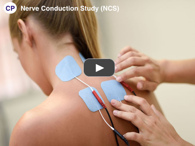
NCV is often used along with an EMG to tell the difference between a nerve disorder and a muscle disorder. NCV detects a problem with the nerve, whereas an EMG detects whether the muscle is working properly in response to the nerve’s stimulus.
Diseases or conditions that may be checked with NCV include:
- Guillain-Barré syndrome; A condition in which the body’s immune system attacks part of the peripheral nervous system. The first symptoms may include weakness or a tingling sensation in the legs.
- Carpal tunnel syndrome; A condition in which the median nerve, which runs from the forearm into the hand, becomes pressed or squeezed at the wrist by enlarged tendons or ligaments. This causes pain and numbness in the fingers.
- Charcot-Marie-Tooth disease; An inherited neurological condition that affects both the motor and sensory nerves. It causes weakness of the foot and lower leg muscles.
- Herniated disk disease; This condition occurs when the fibrous cartilage that surrounds the disks of your vertebrae breaks down. The center of each disk, which contains a gelatinous substance, is forced outward. This places pressure on a spinal nerve and causes pain and damage to the nerve.
- Chronic inflammatory polyneuropathy and neuropathy; These are conditions resulting from diabetes or alcoholism. Symptoms may include numbness or tingling in a single nerve or many nerves at the same time.
- Sciatic nerve problems; There are many causes of sciatic nerve problems. The most common is a bulging or ruptured spinal disk that presses against the roots of the nerve leading to the sciatic nerve. Pain, tingling, or numbness often result.
An NCV procedure may be done on an outpatient basis. Procedures may vary depending on your condition and your doctor’s practices. The NCV is done by a neurologist. This is a doctor who specializes in brain and nerve disorders. A technologist may also do some parts of the test.
The paste used to attach the electrodes will be removed from your skin. After the test, you may return to your previous activities, unless your physician advises you differently. Your physician may instruct you to avoid strenuous activities for the rest of the day.
When your orthotics are a good fit for you, they will not cause knee pain. They will cushion and support your feet while making it easier to maintain good posture. Because no two people have an identical musculoskeletal system, customization is key to obtaining high-quality orthotics.
The “break-in period” can take time. You may have to wear your custom orthotics, identify small problems, and have them reshaped or adjusted gradually. In the end, you should be able to obtain orthotics that are perfectly suited to your body and your lifestyle which relieve pain and help prevent injury.
If your orthotics are not right for you, you may experience knee pain. Orthotics can often change the way the user holds and moves their body while walking or standing, altering weight distribution and foot mechanics. An extreme example is a person wearing high-heeled shoes. Wearing these shoes will change the way they stand. Similarly, using an orthotic insert will lift one part of the foot, changing the person’s entire posture. Even if the change in posture is slight, if done incorrectly, over time it can cause stress and strain above and beyond the original condition.


